Liver is a vital organ that aids in the process of digestion, removal of toxic and waste products from the body, etc. Damage to the liver leads to conditions such as jaundice, hepatitis, cirrhosis, hemochromatosis, Wilson’s disease, non-alcoholic fatty liver disease, cancer, etc. Several factors are responsible for either inducing hepatic injury or worsening the liver damage process. These include chemicals or drugs, oxidative stress, viral infections, excessive consumption of alcohol, accumulation of bile acid, over activation of cells such as hepatocytes, Kupffer cells, fat storing stellate cells and leukocytes present in the liver, etc.
Changes in biochemical markers such as alanine aminotransferase (ALT), alkaline phosphatase (ALP), bilirubin, aspartate transaminase (AST), albumin, gamma glutamyl transpeptidase, extracellular matrix proteins (collagen), transforming growth factor (TGF-β1), etc., are often used to indicate liver damage.
THC’s and curcumin increased the antioxidant defense through free radical scavenging in chloroquine (CQ)-induced toxicity. As THCs are more rapidly metabolized during intestinal absorption than curcumin, they show better and prominent effect in attenuation of CQ-induced lipid peroxidation (Pari and Amali, 2005).
In the study, female wistar rats were administered CQ and THCs for 8 days orally before single administration of CQ and treatment with THC’s followed for 7 more days. THCs showed a significant decrease in the activities of AST, ALP, ALT, levels of bilirubin and lipid peroxidation products, increase in the activity of non-enzymatic antioxidants such as plasma and liver vitamin C and vitamin E, glutathione, increase in the enzymatic antioxidants such as superoxide dismutase, catalase and glutathione peroxidase as compared to CQ-treated control rats. Histopathology revealed near normal appearance of the liver which otherwise showed feathery degeneration and microvesicular type of fatty regeneration, sinusoidal dilation and focal necrosis in CQ-treated rats (Pari and Amali, 2005).

(Adapted from Muthumani and Miltonprabu, 2015)
Pathological changes of the liver such as inflammatory infiltration filling over the sinusoidal vacuolation of hepatocytic nuclei and portal triad with mild inflammation and cell infiltration were reduced in rats treated with THCs (Pari and Amali, 2005).
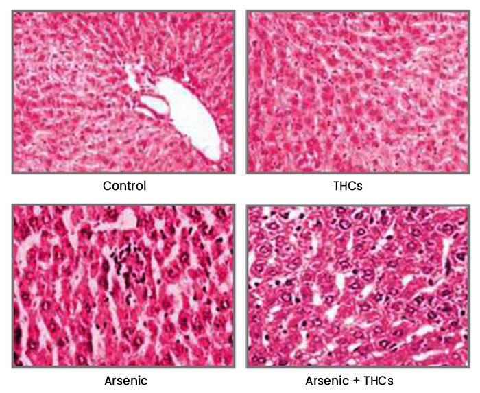
In another study, Muthumani and Miltonprabu (2015) investigated the hepatoprotective effect of THCs over arsenic (a potent hepatotoxin)-induced toxicity in rat liver. Hepatotoxicity was measured by the increased activities of serum hepato-specific enzymes, namely ALT, ALP, AST and bilirubin along with increased elevation of lipid peroxidative markers, TBARS. The results indicated that THCs reduced the levels of liver enzymes including transaminases, ALP, LDH and bilirubin in rats. Treatment with THCs also reduced the oxidative stress, dyslipidemia, mitochondrial damage and improved mitochondrial structure and function in arsenic exposed rat liver.

(Adapted from Muthumani and Miltonprabu, 2015)
Most recently, Ramakrishnan et al. (2017) studied the protective role of THCs on Cd-induced oxidative damage in rats. Tetrahydrocurcuminoids (20, 40 and 80 mg/kg/bw) were orally administered followed by Cd for 4 weeks. Histopathological changes observed in Cd-intoxicated hepatic tissues were minimized on treatment with THCs. The authors concluded that THCs at the dose of 80 mg/kg/bw effectively subdues the Cd-induced toxicity and controls the free radical-induced liver damage in rats.
Protective role of Tetrahydrocurcumin (THC), an active principle of turmeric on chloroquine (CQ)-induced hepatotoxicity in rats
The main objective of the study was to induce hepatotoxicity by CQ and to demonstrate the protective effect of THC in rats treated with CQ. Single oral administration of CQ in female wistar rats (180 – 200 g body weight), significantly increased the activities of serum enzymes namely aspartate transaminase (AST), alanine transaminase (ALT) and alkaline phosphatase (ALP) as well as the levels of bilirubin. Significant alterations of lipids in serum and lipidperoxidation markers as well as hydroperoxides in the plasma and liver were also elevated. The rats administered with THC (80 mg/kg body weight) and curcumin (80 mg/kg body weight) by oral route for 8 days before and 7 days after single administration of CQ showed significantly decreased activities of serum markers and lipids in serum. The level of hydroperoxides was significantly decreased while the non-enzymatic and enzymatic antioxidants were significantly increased upon treatment with THC and curcumin. The study concludes that THC and curcumin exert significant protection against CQ-induced toxicity by their ability to ameliorate lipid peroxidation through free radical scavenging activity. Further, THC has greater effect than curcumin in CQ-induced lipid peroxidation since THC is the rapidly metabolized product of curcumin during absorption in intestine (Pari and Amali, 2005).
Protective role of THC against erythromycin estolate-induced hepatotoxicity
The main objective of this study was to investigate hepatoprotective effect of THC in wistar rats against erythromycin estolate-induced toxicity. Oral administration of THC prevented occurrence of erythromycin estolate-induced liver damage in female wistar rats. The rats treated with erythromycin estolate showed an increase in the level of serum enzymes, bilirubin, cholesterol, triglycerides, phospholipids, free fatty acids and plasma thiobarbituric acid reactive substances (TBARS), whereas they were found to be reduced levels in rats treated with THC and erythromycin estolate. The erythromycin estolate-treated rats may potentiate focal hepatocyte damage and degeneration, which is provoked by the increased production of a high reactive intermediate of erythromycin estolate–nitrosoalkane derivative. The study results conclude that THC is a hepato-stimulant and exerts significant hepatoprotection against erythromycin estolate-induced toxicity (Pari and Murugan, 2004).
